Saturday, January 31, 2009

How to spot Swine Dysentery


Swine dysentery (SD), is caused by a spirochaetal bacterium called Brachyspira hyodysenteriae. This organism causes a severe inflammation of the large intestine with a bloody mucous diarrhoea (i.e. dysentery). 
Disease is common in pigs from 12 to 75kg but severe cases occur occasionally in sows and their sucking piglets. 
SD will survive outside the pig for up to seven weeks in cold moist conditions but it dies out in two days in dry warm environments. 
Spread through the herd is slow, building up in numbers as the dose rate of the causal agent builds up in the environment. Pigs that recover develop a low immunity and rarely suffer from the disease again. 
The high cost of disease is associated with mortality (low), morbidity (high), depression of growth and feed conversion efficiency, and costs of continual in-feed medication.
The incubation period in field cases is normally 7 to 14 days but can be as long as 60 days. Pigs may develop a sub-clinical carrier state initially and then break down with clinical disease when put under stress or when there is a change of feed.

Symptoms

Sows
  • Clinical disease in sows is uncommon unless new disease appears in the herd.
Piglets
  • Severe acute dysentery may occur.
  • Sloppy light brown faeces with or without mucous or blood.
  • Loss of condition.
  • Sows become symptom less carriers. 
Weaners and Growers
The first signs are:-
  • Sloppy diarrhea, which stains the skin under the anus.
  • Initially the diarrhea is light brown and contains jelly-like mucus and becomes watery.
  • Twitching of the tail.
  • Hollowing of the flanks with poor growth.
  • Partial loss of appetite.
  • Slight reddening of the skin.

As the disease progresses:
  • Blood may appear in increasing amounts turning the faeces dark and tarry.
  • The pig rapidly loses condition.
  • Becomes dehydrated.
  • A gaunt appearance with sunken eyes.
  • Sudden death sometimes occurs mainly in heavy finishers.

Causes / Contributing factors

  • Pigs become infected through the ingestion of infected faeces.
  • Spread is by carrier pigs that shed the organism in faeces for long periods.
  • It may enter the farm through the introduction of carrier pigs.
  • Mechanically in infected faeces via equipment, contaminated delivery pipe of feed vehicles, boots or birds.
  • It can be spread by flies, mice, birds and dogs.
  • Stress resulting from change of feed may precipitate.
  • Poor sanitation and wet pens enhance the disease.
  • Overcrowding.
  • It is a major disease in the growing pig but the breeding female can become a carrier for a long period of time and therefore acts as a potential source of infection to other pigs.

Diagnosis

This is based on the history, the clinical picture, post-mortem examinations, laboratory tests on faecal smears and the isolation and identification of S. hyodysenteriae by serological and biochemical tests and DNA analysis. Identification requires specialised procedures which are not available in every laboratory. 
Post-mortem examinations show that the lesions are confined to the large bowel and sometimes the greater curvature of the stomach. 

The disease has to be distinguished from colitis caused by other spirochetes, non-specific colitis, PIA and bloody gut (PHE), acute salmonella infections and heavy infections of the whip worm, trichuris.


How to spot Swine Dysentery

The disease usually affects olders pigs
especially those in the starter-grower stages
 
Brownish Diarrhea
Diarrhea progresses to brownish red
with a much more liquid consistency
 
 Lesions are found in the large intestine
 
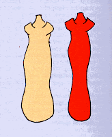
Typical shape of the swine dysentery infected pigs

Friday, January 9, 2009

How to spot Hog Cholera


Pigs affected with hog cholera appear weak, staggering and tend to sit like a dog.


Pigs showing erythema in different areas of the skin


Purplish discoloration of the skin, ears, abdominal part, legs

'Goose-stepping' in hog cholera

Running movement, a nervous sign in hog cholera

Button ulcers in the ceco-colic

Normal-sized spleen with infarcts at the margin (black spots)

Petechial hemorrhages in the mucusa of the urinary bladder

Petechial hemorrhages in the kidney (turkey-egg kidney)

Enlarged and hemorrhagic lymph nodes
Piglets born from a hog cholera-infected sow

Hog Cholera

(From this useful site )

Definition

Hog cholera (HC) is a highly contagious viral disease of swine that occurs in an acute, a subacute, a chronic, or a persistent form. In the acute form, the disease is characterized by high fever, severe depression, multiple superficial and internal hemorrhages, and high morbidity and mortality. In the chronic form, the signs of depression, anorexia, and fever are less severe than in the acute form, and recovery is occasionally seen in mature animals. Transplacental infection with viral strains of low virulence often results in persistently infected piglets, which constitute a major cause of virus dissemination to noninfected farms.

Transmission

The pig is the only natural reservoir of HCV. Blood, tissues, secretions and excretions from an infected animal contain HCV. Transmission occurs mostly by the oral route, though infection can occur through the conjunctiva, mucous membrane, skin abrasion, insemination, and percutaneous blood transfer (e.g., common needle, contaminated instruments). Airborne transmission is not thought to be important in the epizootiology of HC, but such transmission could occur between mechanically ventilated units within close proximity to each other.

Introduction of infected pigs is the principal source of infection in HC-free herds. Farming activities such as auction sales, livestock shows, visits by feed dealers, and rendering trucks are also potential sources of contagion. Feeding of raw or insufficiently cooked garbage is a potent source of HCV. During the warm season, HCV may be carried mechanically by insect vectors that are common to the farm environment. There is no evidence, however, that HCV replicates in invertebrate vectors. Husbandry methods also play an important role in HC transmission. Large breeding units (100 sows) have a higher risk of recycling infection than small herds. In large breeding units where continuous farrowing is practiced, strains of low virulence may be perpetuated indefinitely until the cycle is interrupted by stamping-out procedures and a thorough cleaning and disinfection are carried out.


Incubation Period

The incubation period is usually 3 to 4 days but can range from 2 to 14 days.


Clinical Signs

The clinical signs of HC are determined by the virulence of the strain and the susceptibility of the host pigs. Virulent strains cause the acute form of the disease, whereas strains of low virulence induce a relatively high proportion of chronic infections that may be inapparent or atypical. These strains are also responsible for the "carrier-sow" syndrome from which persistently infected piglets are produced.


Wednesday, January 7, 2009

UN mission investigates Ebola-Reston outbreak in Philippines

From Yahoo News

MANILA (AFP) – Experts from three UN agencies have arrived in the Philippines to investigate an outbreak of the Ebola-Reston virus at two pig farms north of Manila, the World Health Organisation said Wednesday.
Ebola-Reston, which is only found in the Philippines, had been confined to monkeys and the latest outbreak is the first time it has jumped species.

According to the WHO, the strain is not dangerous to humans, unlike the four deadly Ebola subtypes found in Africa.The Food and Agriculture Organisation and the World Organisation for Animal Health are also represented in the mission."The fact that this is the first time that the virus has been found outside monkeys, and the first time ever, worldwide, that it has been found in swine, a food-producing animal, makes this mission particularly important," a WHO statement said.

It added the case had "potential implications for animal and human health and welfare.

"The Philippine government quarantined the affected farms in Santo Nino and Pinagpanaan villages and halted pork exports when the virus was first detected in October.

The UN team was set to work with its Filipino counterparts over 10 days to establish "the source of the virus, its transmission, its virulence and its natural habitat, in order to provide appropriate guidance for animal and human health protection.

"Preliminary results are expected in a few weeks at the earliest," said the statement.

Local authorities have been unable to find any sign of the virus among farmhands or slaughterhouse workers who handled the pigs.

Ebola-Reston was first detected in 1989 in laboratory monkeys sent from the Philippines to Reston in the United States.